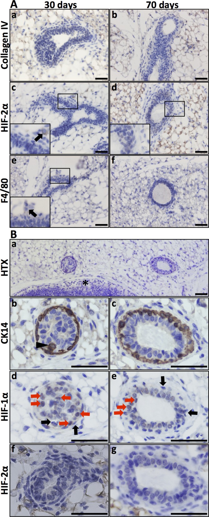
Fig 2. HIF-1α and HIF-2α expression in the virgin mammary gland.

A. Virgin mammary glands (30 and 70 days old) showed no conspicuous basal membrane, as visualised by collagen IV IHC (a, b). There was also no detectable expression of HIF-2α in the epithelial cells (c, d). Macrophages (F4/80 positive) were few. In panel c, a single HIF-2α-positive cell was detected, and the adjacent F4/80 IHC section (e) suggested that this cell is a macrophage. B. Expression of HIF-1α in mammary epithelium in the 70-day-old virgin mouse. Top panel, orientation slide with haematoxylin (HTX) staining, 20x obj. *lymph node. Panels b, d, f. Cross-section of a developing duct close to the invading tip at a stage where the lumen is not yet evacuated, 40x obj. Panels c, e, g. Cross-section of a less mature part of a duct, 40x obj. CK14-expressing cells (marker of basal mammary epithelial cells) can be seen in more than one cell layer (panels b and c, arrow-head). At this stage, the lumen is evacuated, but there is still more than one layer of epithelial cells. HIF-1α IHC on the adjacent sections (panels d, e) showing nuclear expression in non-basal epithelial cells (highlighted by red arrows). Basal (CK14 positive) epithelial cells did not express HIF-1α (black arrows). Mammary epithelial expression of HIF-2α was not detected at these developmental stages. Size bars: 50 μm.
