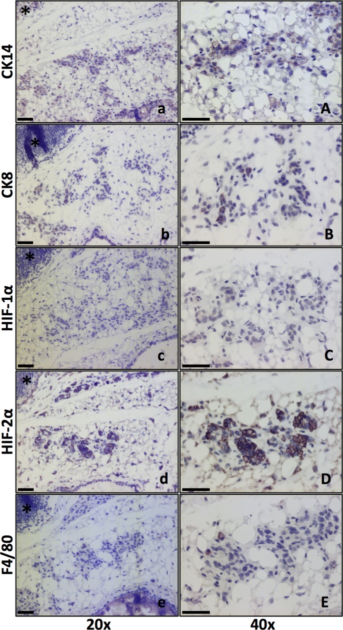
Fig 5. HIF-alpha expression in involuting glands five days post weaning.

Size bars: 50 μm. * marks the lymph node for orientation. 20x and 40x lenses as indicated. a, A. CK14 marks the basal mammary epithelial cells and stem/progenitor cells. b, B. CK8 positivity shows the luminal mammary epithelial cells. c, C. HIF-1α IHC showed weak or little positivity in mammary epithelial cells at this stage. d, D. HIF-2α positive cells were found in the clusters of epithelial cells. e, E. Macrophage (F4/80 positive) infiltration has begun by five days post weaning. F4/80 positive cells were apparently fewer than HIF-2α positive cells.
