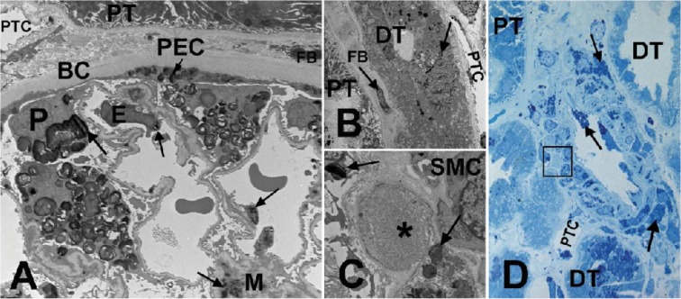
Fig 4. Kidney biopsy images from a male patient with Fabry disease.
(A) EM of a glomerulus. Arrows show GL-3 inclusions in podocytes (P), endothelial cells (E), mesangial cells (M), and parietal epithelial cells (PEC). (B) EM of a distal tubule (DT) with GL-3 inclusions (arrows) accumulated in its epithelial cells, in contrast to the adjacent proximal tubule (PT) with no obvious GL-3 inclusion. (C) EM close-up of the square shown in (D), displaying early Fabry arteriopathy (asterisk) focally replacing smooth muscle cells (SMC) of an arteriolar wall. Arrows show GL-3 inclusions in adjacent smooth muscle cells. (D) Methylene blue/Azure II (Richardson’s) stained semi-thin section of an arteriole. Note that the arteriopathy shown in (C) with higher magnification EM is not easily identifiable by LM (square). Arrows show GL-3 inclusions in endothelial cells and smooth muscle cells of the artery. BC = Bowman's capsule; DT = distal tubule; E = endothelial cells; EM = electron microscopy; FB = fibroblast; GL-3 = globotriaosylceramide; LM = light microscopy; M = mesangial cells; P = podocytes; PEC = parietal epithelial cells; PT = proximal tubule; PTC = peritubular capillary; SMC = smooth muscle cells. EM measurements of GL-3 deposition in glomerular cell types are shown in Table 4 [16]. Unbiased morphometric EM estimates of GL-3 in the three glomerular cell types including Vv (Inc/PC), Vv(Inc/Endo), and Vv(Inc/Mes) [16,27], as well as podocyte foot process width and percentage glomerular capillary endothelial fenestration [16,26,27] are tabulated in Table 4.

