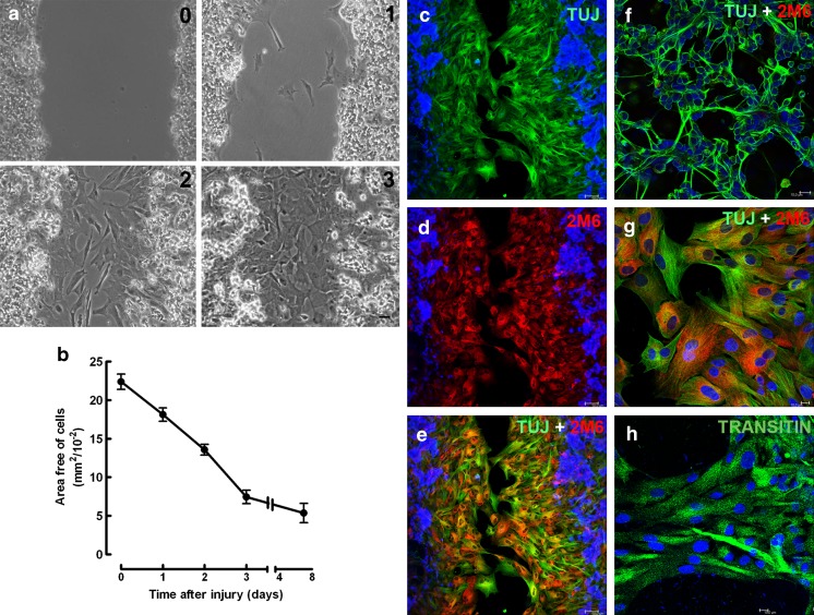
Fig. 1.
Time course of glial growth over the scratched area in chick embryo retinal monolayer cultures. Retinal cell cultures from 8-day-old chick embryos maintained for 7 days (E8C7) were scratched, the medium changed, and cultures cultivated for several days. a Phase contrast micrographs showing the scratched area 0, 1, 2, or 3 days after the scratch of the cultures. b Quantification of the area free of cells as a function of time after the scratch. On the day indicated, 3 to 10 fields of 0.325 mm2 per culture were photographed under phase contrast illumination each day and the growth of cells estimated as the decrease in the area devoid of cells. c–e Three days after the scratch of the monolayer, immunocytochemistry against the neuronal marker beta-tubulin III (green) and the glial antigen 2 M6 (red) was performed. Cell nuclei were stained with DAPI (blue). f Photomicrograph of the cultures showing that neurons are beta-tubulin III positive, but 2M6 negative. g Confocal micrograph of the scratched area showing that 2 M6-positive glial cells (red) are also labeled with TUJ1 antiserum (green). h Photomicrograph of glial cells of the scratched area labeled for transitin (green). Quantitative data represent the mean ± SEM in mm2/10−2 of three to five separate experiments. Bar = 50 μm (a, c, d, e) and 10 μm (f, g, h)

