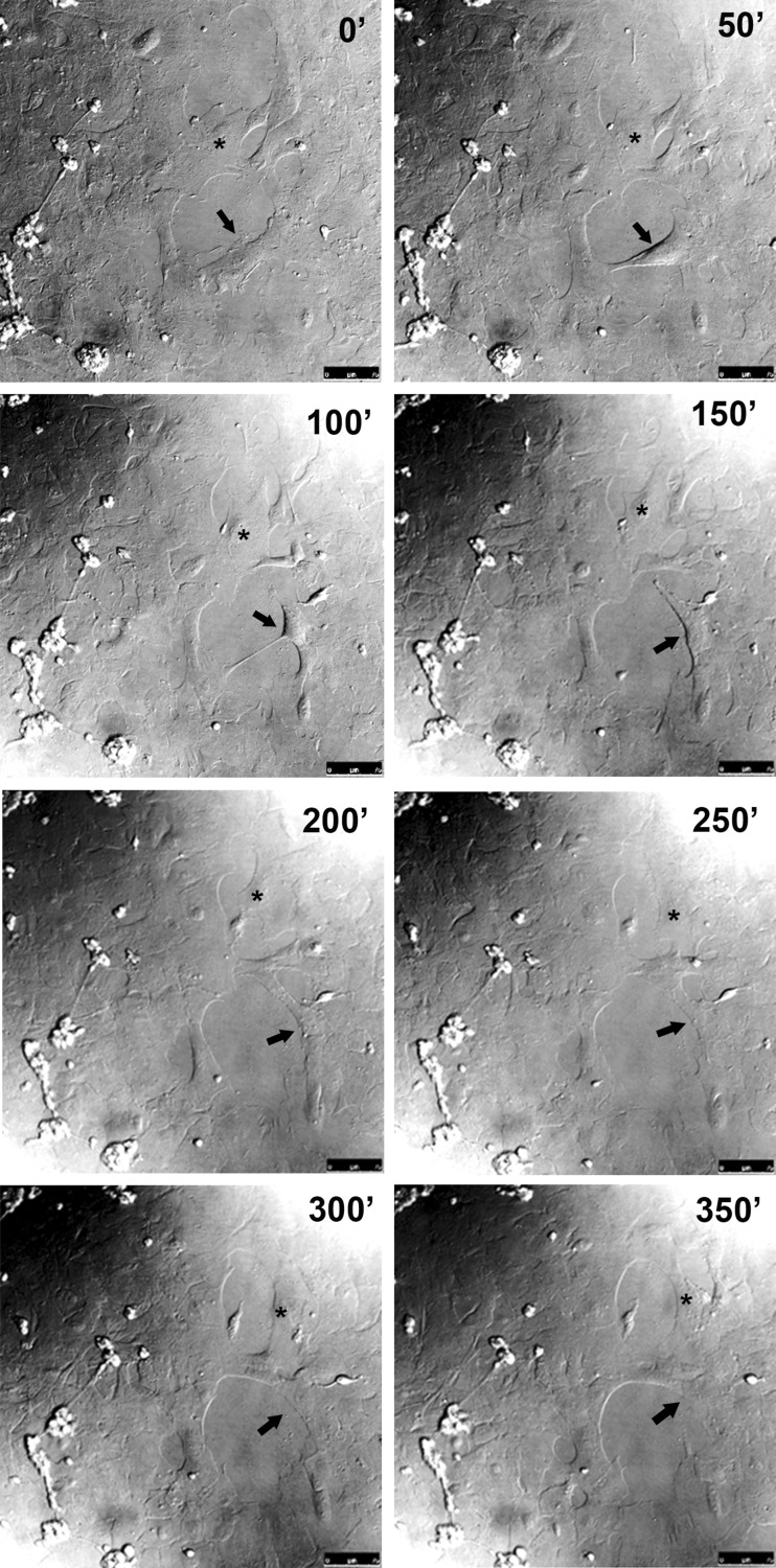
Fig. 2.
Glial cell migration and division in the scratched area of retinal monolayer cultures. Retinal cell cultures at E8C7 were scratched, and after 3 days, culture medium was changed to MEM buffered with 25 mM HEPES (pH 7.4) plus serum and antibiotics and cultures mounted on a confocal microscope with a culture chamber. Cultures were photographed at the indicated time periods under differential interference contrast illumination using ×20 objective. A migrating (asterisk) and a dividing (arrow) glial cell are shown. Bar = 75 μm

