Fig. 6.
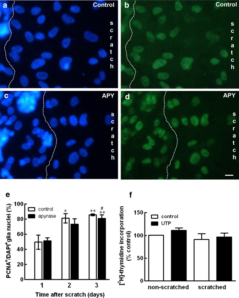
Effect of apyrase on glial cell proliferation at the edge of the scratched area of the cultures. Retinal cultures were scratched and incubated in the absence (a, b) or presence (c, d) of 2.5 U/mL of apyrase for 3 days. Cell nuclei were stained with DAPI (a, c) or with antiserum against PCNA (b, d). e Ratio of PCNA+/DAPI+ glial nuclei determined in micrographs of the edge of the scratch. The area of neurons was excluded with a dashed line as shown in a–d, and glia nuclei were counted at the area between neurons and the center of the scratch. PCNA+/DAPI+ ratios were determined 1, 2, or 3 days after scratching the cultures. f Cultures at E8C8 that were scratched or not at E8C7 were incubated with 100 μM UTP for 24 h and the incorporation of [3H]-thymidine determined as described in the “Materials and methods” section. Data were expressed as % control of non-scratched cultures and represent the mean ± SEM of three to six separate experiments performed in duplicate. Bar = 10 μm

