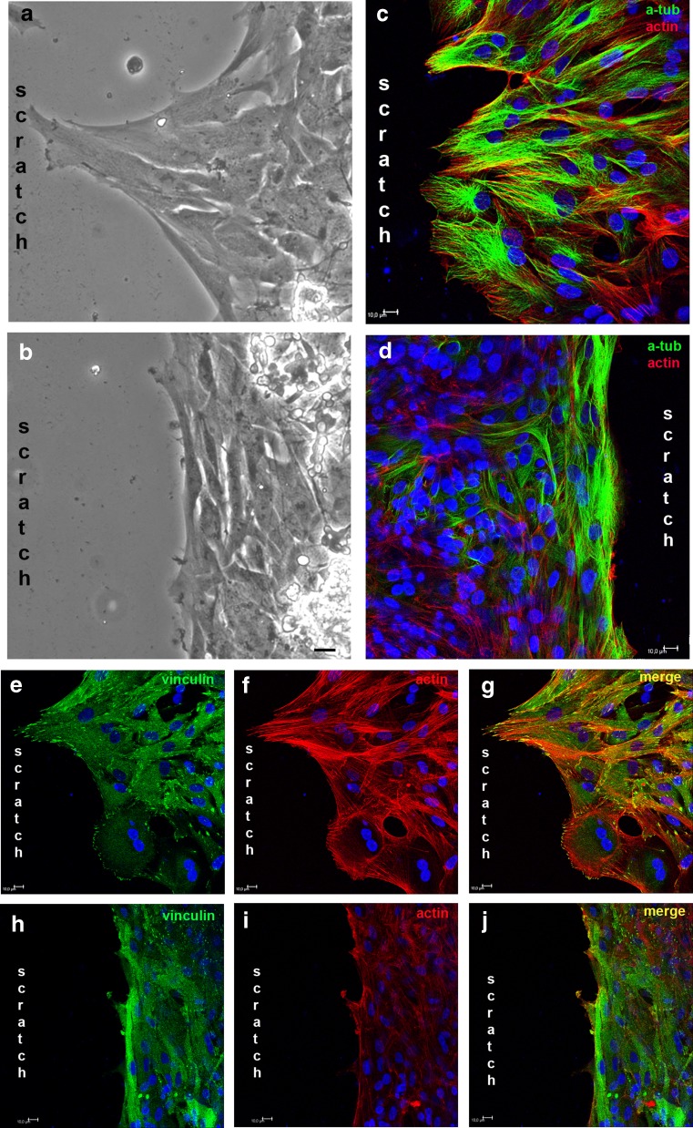
Fig. 9.
Effect of apyrase on the morphology and cytoskeletal arrangement of glial cells at the edge of the scratched area. Cultures at E8C7 were scratched and incubated with 2.5 U/mL of apyrase for 3 days. Then, cultures were fixed and incubated with anti-α-tubulin or anti-vinculin. Phalloidin-Alexa 568 was used to label actin filaments. a, b Micrographs photographed under phase contrast illumination of control and apyrase-treated cultures showing the morphology of glial cells at the border of the scratched area. c, d Alfa-tubulin and actin labeling in control (c) and apyrase-treated (d) cultures. e, f Labeling against vinculin (green) and actin (red) in control cultures. h, i Labeling against vinculin (green) and actin (red) in apyrase-treated cultures. Merged figures are shown in (g) and (h). Bar = 20 μm (a, b) and 10 μm (c–j)

