
Figure 4.
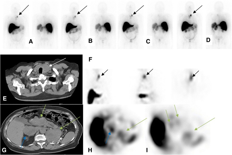
Patient with metastatic renal cell carcinoma post left nephrectomy. Patient received 5 mg of 111In hu-J591. Delayed WB images (day 6 or 7; cycles 1 to 4) show uptake in left neck (A to D, respectively; black arrows) corresponding to the left paratracheal nodal mass seen on CT image (E) (white arrow). This is also seen in the coronal, sagittal, and axial SPECT images (respectively in (F)). Uptake was also seen corresponding to the right adrenal mass (blue arrow), pancreatic head mass (short green arrows), and left renal bed mass (long green arrow) (CT image (G) and axial SPECT images (H) and (I)).