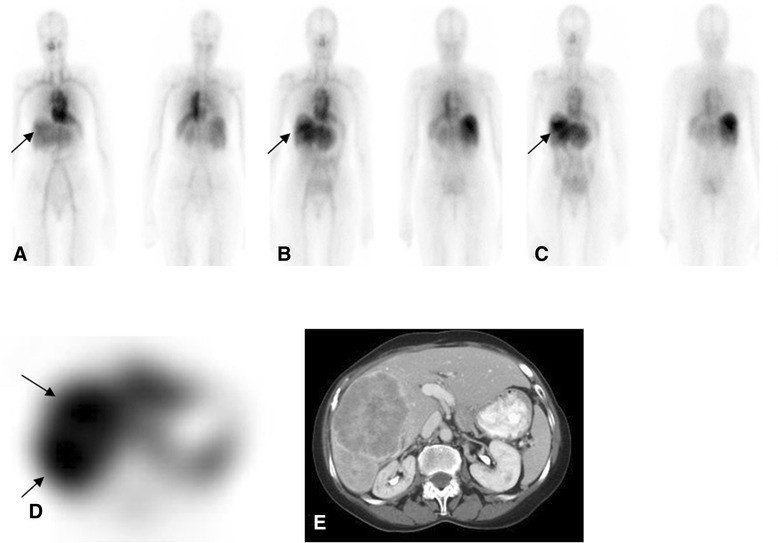
Figure 5.

Female with metastatic hepatocellular carcinoma. Anterior and posterior WB images from day 0, day 3, and day 6 ((A-C), respectively) show heterogeneous activity in the liver steadily increasing in later images (C), more clearly seen on SPECT (D), corresponding to the progressive liver disease seen on CT images (E).
