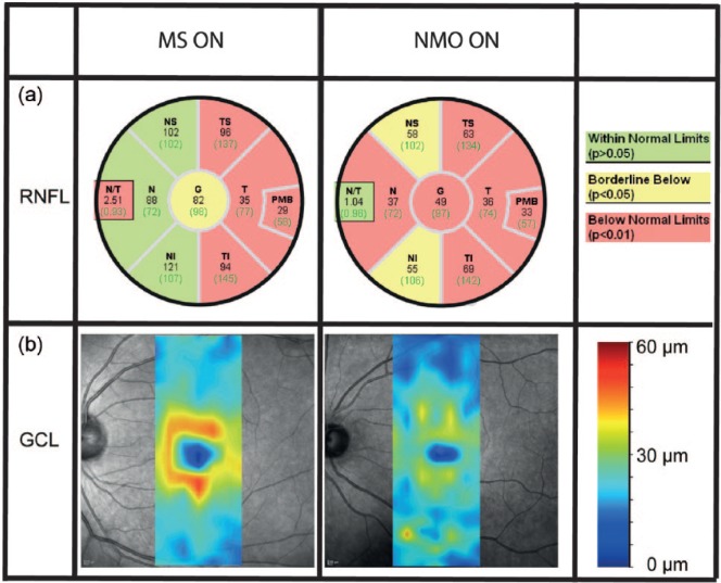
Figure 2.
Typical differences in retinal damage between NMO-ON and MS-ON.
(a) RNFL thickness values for different locations of the peripapillary ring scans including comparison to a healthy reference group. (b) Thickness map of the retinal GCL, derived with help of a semiautomatic segmentation software. The NMO-ON patient shows more severe thinning both in the RNFL and GCL.
MS: multiple sclerosis; ON: optic neuritis; NMO: neuromyelitis optica; RNFL: retinal nerve fiber layer; GCL: ganglion cell layer.

