Figure 5.

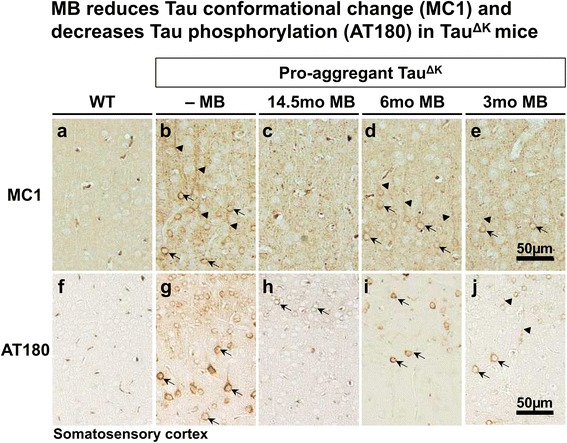
Histological analysis of conformationally changed and phosphorylated Tau in MB-treated mice. (a-e) MC1 immunoreactivity (epitope 5-15 + 312-322) indicates a pathological Tau conformation. MC1 positive neurons are prominent in somatosensory cortex (SSCx) of untreated TauΔK mice with missorting of Tau to cell soma (arrows) and apical dendrites (arrowheads). In contrast MB treatment, especially preventive MB treatment for 14.5mo, clearly reduces MC1 immunoreactivity in TauΔK mice. (f-j) Histological analysis of phosphorylated Tau using the AT180 antibody (dual phosphorylation epitope pThr231 + pSer235). Untreated TauΔK mice display a massive mislocalization of phosphorylated Tau to cell soma (arrows) and apical dendrites (arrowheads) of SSCx neurons, whereas MB treatment diminishes the extent of AT180 phosphorylation. Scale bar: 50 μm.
