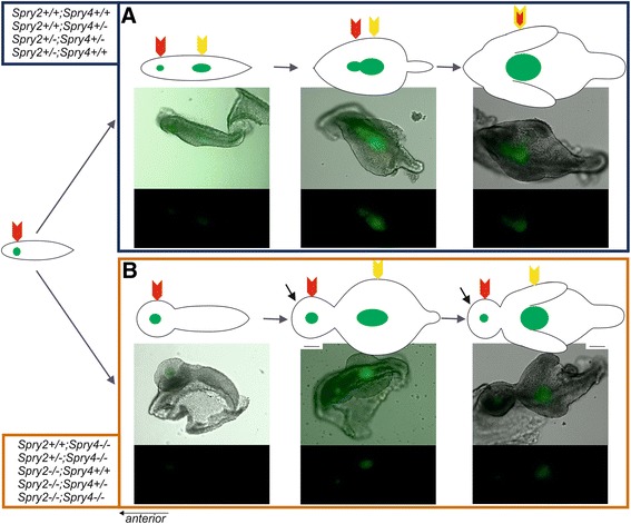
Figure 1.

Physiological and pathological tooth formation in dissociated epithelia of mouse lower cheek region. (A) In samples with higher Spry2;Spry4 gene dosages (Spry2+/+;Spry4+/+, Spry2+/+;Spry4+/−, Spry2+/−; Spry4+/−, Spry2+/−; Spry4+/+), R2 Shh signaling domain (red arrow) merges with early M1 Shh signaling domain (yellow arrow) to form a typical elongated pEK (double yellow-red arrow) of the germ of M1. (B) In samples with lower Spry2;Spry4 gene dosages (Spry2+/+; Spry4−/−, Spry2+/−; Spry4−/−, Spry2−/−; Spry4+/+, Spry2−/−; Spry4+/−, Spry2−/−; Spry4−/−), R2 signaling domain (red arrow) persists in front of M1 signaling domain (yellow arrow) and became a signaling center of supernumerary tooth primordium (black arrow). Anterior direction is on the left side (the scale bar is 100 μm).
