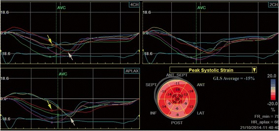
Figure 4.

Left ventricular longitudinal strain measured by speckle tracking echocardiography in a patient with severe aortic stenosis and chest pain. A nonuniform reduction of longitudinal deformation can be observed, with reduced values of peak systolic strain in the basal segments of the interventricular septum (yellow arrows) and post-systolic shortening in mid and basal segments of the lateral wall (white arrows). Coronary angiography revealed a calcified left main stenosis (80%) extended to the origin of the circumflex artery and a hypoplastic right coronary artery.
