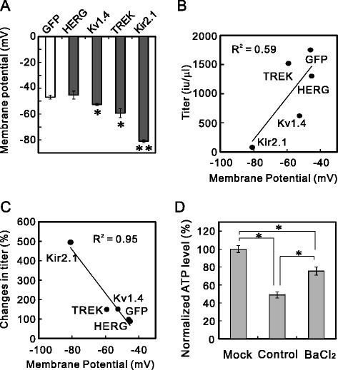
Figure 8.

Correlation between the resting membrane potential and lentiviral titer. (A) Resting membrane potentials of K+-channel-expressing cells. 293T cells were transfected with plasmid Lv-GFP, Lv-HERG, Lv-Kv1.4, Lv-TREK, or Lv-Kir2.1. The membrane potentials of these K+-channel-expressing cells were measured in the whole-cell current-clamp mode 24 h after transfection (*p < 0.05, **p < 0.000001 vs Lv-GFP; ANOVA followed by Student’s t test, n = 5). (B) Correlations between membrane potential and the titers of the lentiviral vectors. The titers of the unconcentrated lentiviral vectors encoding GFP, HERG, Kv1.4, TREK, and Kir2.1, prepared without blockers, tended to correlate with the membrane potentials of the 293T cells expressing these channels. Linear regression was used to correlate the data (R 2 = 0.59). (C) Correlation between membrane potential and the increase in the lentiviral vector titer. The membrane potentials of 293T cells expressing GFP, HERG, Kv1.4, TREK, or Kir2.1 correlated well with the percentage change in the lentiviral titer after the addition of the blockers (R 2 = 0.95). (D) Reduction in ATP level and its restoration by Ba2+. The ATP levels in 293T cells were measured 48 h after transfection, without harvesting the lentiviral vectors. The ATP levels were normalized to that of the mock-transfected cells (n = 4, p < 0.00001; ANOVA followed by Student’s t test).
