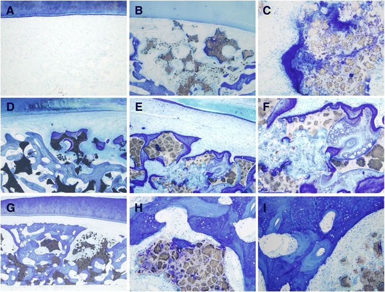
Figure 9.

Histological sections stained by Toluidine blue of the alveolar bone defects at 12 weeks after implantation. (A) No bone regeneration was detected at the alveolar bone control group. A small amount of new bone could be seen in the β-TCP group, with some osteoid formation in the periphery and center of the β-TCP scaffold (B, C). The PDLSCs/β-TCP group showed more new alveolar bone formation, with numerous small bone trusses or trabeculae interconnected with each other (D-F). A maximal and robust bone formation was presented in the hOPG-PDLSCs/β-TCP group. Osteoblastic cells were lining the surface of newly formed bone. Osseous maturation had increased, with osteon formation and increased bone density (G-I). Magnifications: 40× (A, B, D, G), 100× (E), and 400× (C, F, H, I). β-TCP, beta-tricalcium phosphate; hOPG, human osteoprotegerin; PDLSC, periodontal ligament stem cell.
