
Figure 1.
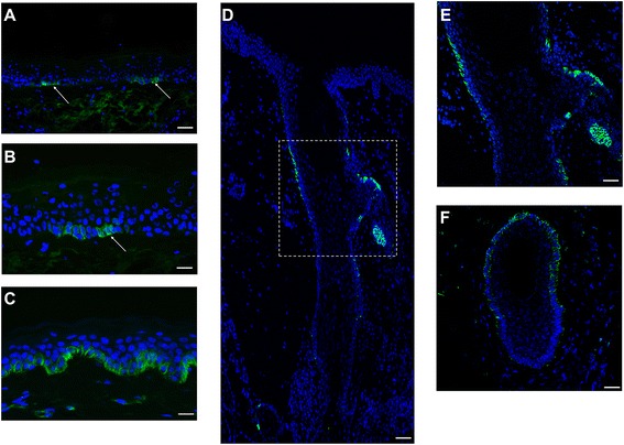
Localization of ABCG2 in human interfollicular skin and hair follicle by immunofluorescence staining of human interfollicular epidermis and hair follicle with anti-ABCG2 antibody (clone BXP-21). Bright green cell membrane staining of ABCG2 was seen in clusters of small cells of basal layer keratinocytes (white arrow) in human skin (A, B) and hair follicle (D, E, F), whereas suprabasal keratinocytes were unstained stained. (B) High magnification of (A). (E) High magnification of (D). (C) Human interfollicular skin was stained with anti-keratin15 antibody (clone LHK15), and most basal cells were positively stained (green) on their cell membrane. Cell nuclei were counterstained with 4′-6-diamidino-2-phenylindole DAPI: (blue). Original magnification: A, D, F, ×10; and B, C, E, ×20. Scale Bars: A, D, F, 50 μm; and B, C, E, 50 μm.