Fig. 3.
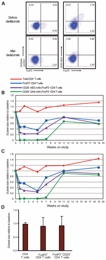
CD25 blockade in vivo depletes systemic Tregs in cancer patients. Peripheral blood samples obtained from patients before and at various times after a single infusion of CD25 mAb daclizumab were analyzed by flow cytometry. (A) Representative data from one patient comparing baseline (before) to 5 weeks after daclizumab. (B and C) Relative changes in the fraction (B) or absolute counts (C) of various T cell subsets over time, shown as the means for all patients normalized to individual baseline values. Daclizumab was given on week 0. Red diamonds, total CD4+ T cells; blue squares, FoxP3+ CD4 T cells; purple triangles, CD25+ FoxP3+ CD4 T cells as identified by the nonblocked monitoring CD25 mAb 4E3; green circles, CD25+ FoxP3+ CD4 T cells as identified by the monitoring CD25 mAb 2A3, which is blocked by daclizumab. For total FoxP3+ CD4 T cells and CD25+ (4E3) FoxP3+ CD4 T cells, P < 0.001 at weeks 1, 2, and 5; for CD25+ (2A3) FoxP3+ CD4 T cells, P < 0.001 at weeks 1, 2, 5, and 7 (statistical details are provided in table S3). (D) Peptide vaccination of breast cancer patients without daclizumab administration does not alter Tregs. Peripheral blood T cell populations were analyzed as fractions before and after vaccination by flow cytometry in a cohort of patients (n = 7) with metastatic breast cancer on a previously described clinical trial (29). Nonblocked mAb 4E3 was used to evaluate CD25. Data are shown as the means of all patients normalized to individual baseline values with error bars representing SD. P > 0.05, comparing Treg fractions at the time of the second or third vaccine to baseline. Similar data were obtained if T cell populations were analyzed as absolute counts.

