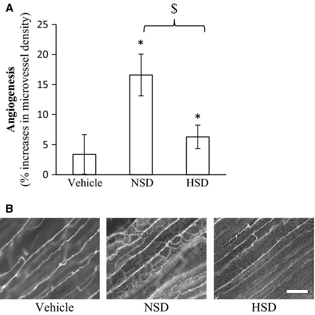
Figure 3.

EPC transplantation with electrically stimulated angiogenesis. Vehicle or 6000 EPCs from NSD or HSD donor rats were injected intravenously through the tail vein of HSD SD donor rats receiving electrical stimulation. Angiogenesis was determined in the tibialis anterior following EPC transplantation. Data expressed as mean ± SE. *Significantly increased in microvessel density between the stimulated and unstimulated limb. $Significantly different between NSD and HSD (P ≤ 0.05, n = 9). (B) Representative images of stimulated muscle, bar = 50 μm.
