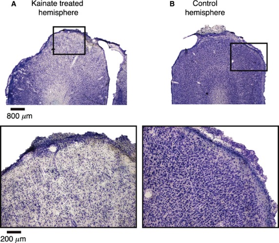
Figure 2.

Effect of kainic acid on the cortex. (A) Single 50 μm thick slice of area 17, stained for Nissl substance, using classical histological techniques, following topical application of kainic acid to the cortical surface in vivo. Top image is at lower magnification and demonstrates the excitotoxic ablation of the affected area as indicated by the decrease in the density of the Nissl stain in the superior portion relative to the lateral and medial extents of the cortex. The bottom image is of the region of the top image enclosed by the black box, which was most affected, viewed under higher magnification to further demonstrate the sparseness of Nissl bodies. (B) As with A, but image of the contralateral cortex of the same animal, on which no kainic acid was applied. The density of Nissl bodies relative to A indicates the extent and efficacy of the ablation.
