
Figure 8.
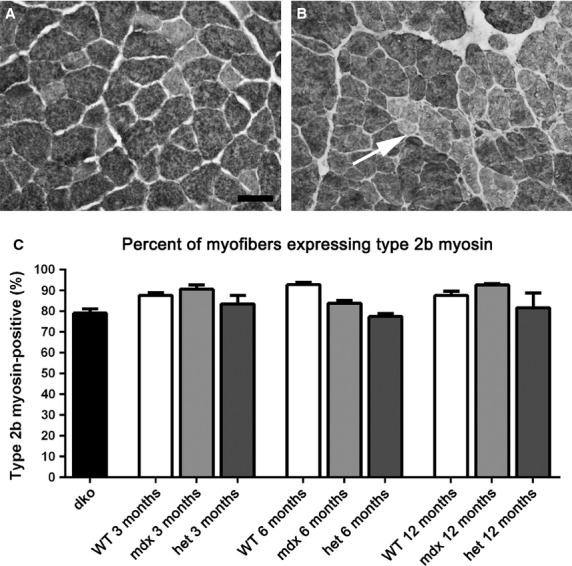
Morphometric analysis of type IIb myofiber density in WT, mdx, mdx:utrophin+/− (het), and dko triceps muscles. (A) The vast majority of the myofibers in the WT triceps muscles were IIb-positive. WT muscle at 6 months is shown. (B) At 6 months in the mdx:utrophin+/− (het) muscles, groups of type IIb negative myofibers were seen indicative of fiber type grouping. Bar is 50 μm. (C) Morphometric analysis of the density of IIb myofibers showed no significant differences between any of the age-matched genotypes.