
Figure 11.
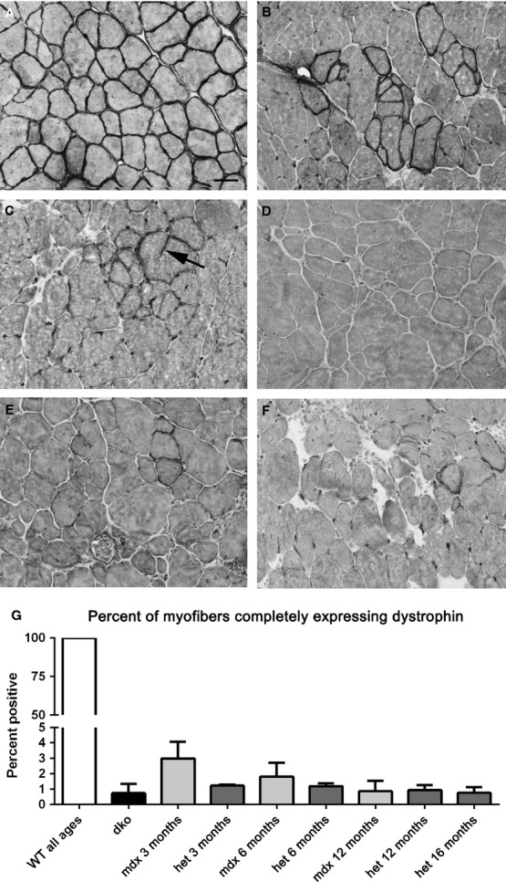
Dystrophin immunostaining in WT, mdx, mdx:utrophin+/− (het), and dko triceps muscles. (A) All the myofibers in WT muscles express dystrophin at their sarcolemmas. (B) mdx muscle at 6 months showing a large cluster of revertant fibers. (C) mdx:utrophin+/− (het) muscle at 6 months. Arrow points to a cytoplasmic extension of dystrophin. (D) mdx:utrophin+/− (het) at 12 months. (E) mdx:utrophin+/− (het) muscle at 16 months. (F) dko at 2 months. (G) Morphometric analysis of revertant fiber density as a percent of all myofibers. There were no significant differences between any of the dystrophic genotypes. Bar is 20 μm.