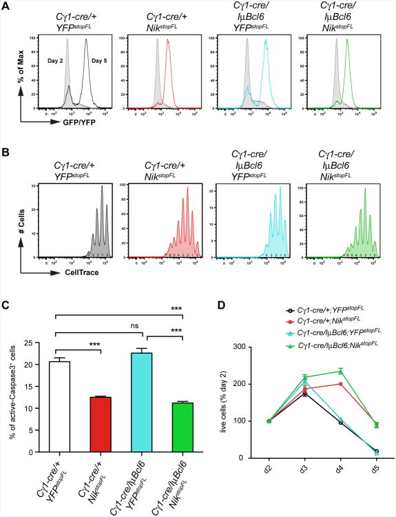
Figure 3. Enhanced Cellular Proliferation and Survival as A Result of Constitutive NIK Expression.
(A) Cre-mediated recombination efficiency in purified B cells from mice of the indicated genotypes, cultured in vitro in the presence of anti-CD40 plus IL4, measured by expression of reporter genes at day 2 and day 5 of the culture.
(B) Proliferation of in vitro cultured B cells treated as in (A), measured by CellTrace dilution of reporter-positive cells at day 5 of the culture. The numbers under CellTrace peaks indicate the number of cell divisions.
(C) Frequency of apoptotic cells, at day 5, within in vitro cultured B cells treated as in (A), measured by active Caspase3 staining.
(D) Relative number of live cells at the indicated time points in in vitro culture of splenic B cells treated as in (A), normalized to day 2.
Data in (A-D) are representative of two independent experiments performed in triplicate; data in (C) are shown as mean ± SEM of triplicates; data in (D) are shown as mean ± standard deviation (SD) of triplicates.

