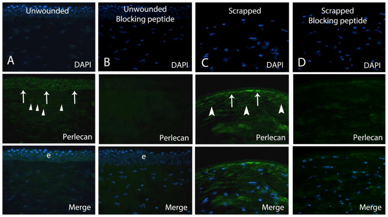
Figure 1.
Representative immunohistochemistry for perlecan in human corneas. Column A. Perlecan protein was noted in the epithelium and epithelial basement membrane (arrows) and at low levels in keratocytes (arrowheads) in unwounded control corneas. Column B. Negative control in unwounded cornea with pre-absorption with blocking peptide shows specific blocking of antigen-antibody interaction. Column C. In all corneas at 30 minutes after epithelial scrape, strong perlecan immunoreactivity was noted in keratocytes (arrowheads), as shown in this representative section from one cornea, including in keratocytes in the early stages of apoptosis that label with the terminal deoxynucleotidyl transferase (TdT)-mediated dUTP nick end labeling (TUNEL) assay (not shown). Staining near the stromal surface (arrows) could represent BM that was not completely removed by scrape or expression in keratocytes undergoing apoptosis—which cannot be distinguished at this level of magnification. Column D. Pre-absorption with blocking peptide shows specific blocking of antigen-antibody interaction in the negative control cornea after epithelial scrape. Blue is DAPI staining of cell nuclei. e is epithelium. Magnification 400x.

