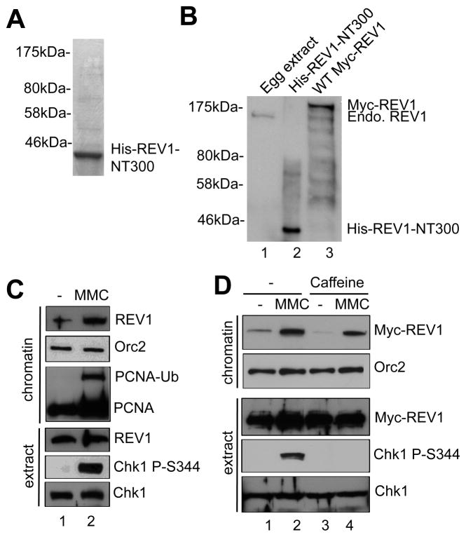
Fig. 2.
REV1 preferentially associates with MMC-damaged chromatin. (A) Purified His-REV1-NT300 was examined via SDS-PAGE and Coomassie blue staining. Molecular weight markers were also labeled. (B) Xenopus egg extract, recombinant His-REV1-NT300 and SP6 TnT expressed WT Myc-REV1 were examined via immunoblotting analysis using anti-REV1 antibodies. “Endo. REV1” represents endogenous REV1. (C) MMC was incubated in egg extracts supplemented with sperm chromatin. After 1-hr incubation, chromatin fractions and total extract were isolated and examined via immunoblotting analysis. “PCNA-Ub” denotes monoubiquitinated PCNA. (D) WT Myc-REV1 was incubated in egg extracts at a similar concentration as the endogenous REV1, which was followed by addition of MMC and Caffeine. After 1-hr incubation, chromatin fractions and total extract were analyzed via immunoblotting analysis as indicated. Orc2 was used as a loading control.

