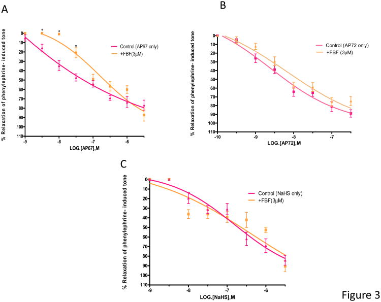
Figure 3.
Concentration-dependant relaxation of phenylephrine-induced tone in isolated bovine ciliary artery by H2S donors (A) AP67, (B) AP72 and (C) NaHS: control, and in the presence of flurbiprofen (FBF, 3 μM). FBF blocked relaxations induced by lower concentrations of AP67 but not those elicited by AP72 and NaHS. Vertical bars represent means ± S.E.M.; n=6--36. *p<0.05, significantly different from control.

