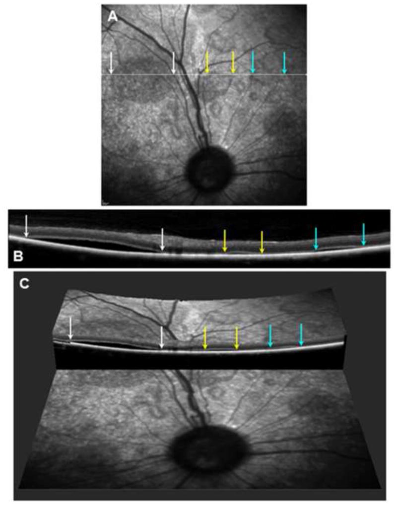
Figure 2.

(A) Scanning laser ophthalmoscopic image of the fundus from a dog exhibiting multiple localized serous detachments. A scan line for the OCT image shown in (B) is shown in white. The edges of 3 of these lesions traversed by the scan line are indicated by colored arrows. (B) An OCT cross-section image of the retina from along the scan line shown in (A) with the edges of the same 3 lesions indicated by the same colored arrows. (C) A 3-dimensional reconstruction of the same area of the retina made by combining multiple OCT images showing the same 3 serous detachments.
