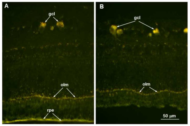
Figure 9.

Fluorescence micrographs of an unstained cryostat section of the retina from a region where the retina remained attached to the RPE (A) and a region within one of the localized retina detachments (B). In both areas the disease-related autofluorescent material was present primarily in the ganglion cells (gcl) and outer limiting membrane (olm). The retinal pigment epithelium (rpe) also contained autofluorescent material with similar spectral properties, but this likely consisted primarily of normal age pigment since similar RPE autofluorescence is present in genetically normal dogs. Scale bar in (B) indicates magnification for both micrographs.
