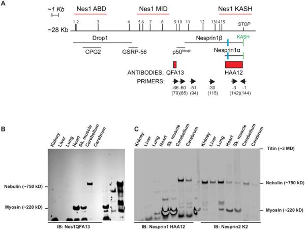
Figure 1.
A) Localization of unique ARCA1 mutations (1 to 15), hybridization probes (red lines), antibody epitopes (red rectangles), floxed exon in ActinCreNesprin1Δ/Δ mice (blue rectangle), primers (forward and reverse arrows above corresponding exon number) and known Nesprin1 isoforms relative to Nesprin1 giant transcript XM_006512463.1 used as a reference in this work. The reverse exon numbering system used in (Zhang et al., 2009a) that simplifies exon labeling was adopted in this work. The classical numbering of corresponding exons of reference transcript XM_006512463.1 is also indicated between brackets. B, C) Autoradiogram of mouse tissue lysates (40µg) immunobloted with Nesprin1 QFA13 (B). Note the high molecular weight band expressed in the cerebellum. A single membrane with duplicate loading of the same tissues was immunobloted with Nesprin1 HAA12 (C, left panel) and Nesprin2K2 (C, right panel) and imaged quantitatively. Note the CNS-specific expression pattern of Nesprin1giant by comparison to Nesprin2 giant. Molecular weight markers correspond to large proteins abundantly expressed skeletal muscle used as molecular weight rulers (Fig.S2).

