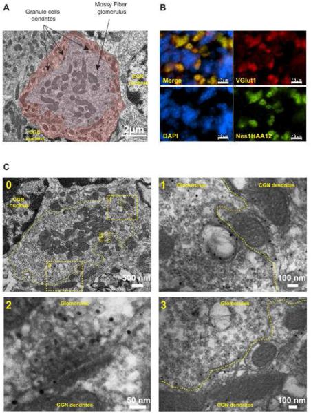
Figure 5.
A) Transmission electron microscopy of a mossy fiber glomerulus within the GCL of a P30 cerebellum from a C57/Bl6 mouse. The glomerulus is pseudocolored in light brown and the adjacent CGN dendrites are pseudocolored in dark brown. B) Coimmunolabeling of the GCL with Vglut1 and Nes1HAA12. C) Immunogold electron microscopy of cerebellar glomeruli and surrounding CGN dendrites. A single glomerulus is delineated by a dashed line in the low magnification image (0). 1 and 3: higher magnification of two regions revealing the colocalization of KLNes1g with a significant pool of synaptic vesicles and with dendritic membranes. 2: Higher magnification showing KLNes1g immunoreactivity within synaptic vesicles at a release site.

