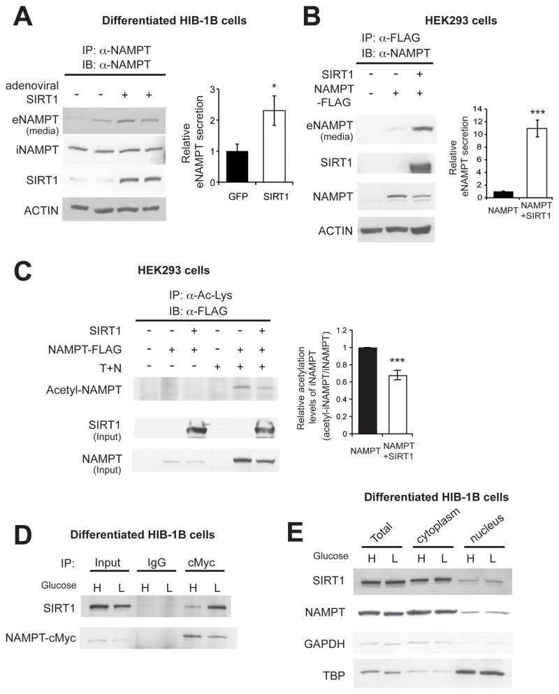
Figure 3.
SIRT1 promotes eNAMPT secretion by deacetylating iNAMPT. (A and B) SIRT1 overexpression promotes eNAMPT secretion in differentiated HIB-1B cells (A) and HEK293 cells (B). Levels of eNAMPT secretion were calculated as described in Materials and Methods. The right panels represent average values from three independent experiments. Each value is normalized to those of GFP- or NAMPT-expressing cells. (C) SIRT1 overexpression decreases iNAMPT acetylation levels in HEK293 cells. Cells were incubated in the absence or presence of 5 μM Trichostatin A (T) and 5 mM nicotinamide (N) overnight prior to cell lysis. Acetylated iNAMPT levels are normalized to total iNAMPT levels. The right panel represents iNAMPT acetylation levels normalized to total iNAMPT levels in each condition (n=3). (D) Interaction between SIRT1 and iNAMPT in differentiated HIB-1B cells. (E) Subcellular localization of SIRT1 and iNAMPT in differentiated HIB-1B cells. GAPDH and TBP were examined as representative cytoplasmic and nuclear proteins, respectively. Cells were incubated with media containing high glucose (H, 25mM) or low glucose (L, 5 mM) overnight (D) or for 3 hr (E). Data were analyzed by the Student’s t test. All values are presented as mean ± SEM. *p ≤ 0.05; **p ≤ 0.01; ***p ≤ 0.001.

