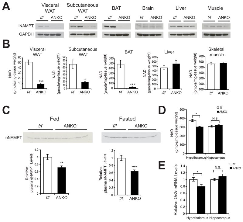
Figure 5.
ANKO mice exhibit reduced plasma eNAMPT levels and defects in NAD+ biosynthesis not only in adipose tissue but also in the hypothalamus. (A) iNAMPT levels in visceral white adipose tissue (WAT), subcutaneous WAT, brown adipose tissue (BAT), brain, liver, and muscle from 2-month-old female Namptflox/flox (f/f) and ANKO mice. (B) Tissue NAD+ levels in visceral WAT, subcutaneous WAT, BAT, liver, and skeletal muscle from 3–4-month-old female Namptflox/flox and ANKO mice (n=4–8). (C) Plasma eNAMPT levels in 5–6-month-old female Namptflox/flox and ANKO mice. Plasma was collected from mice fed or fasted for 48 hr. Bottom panels show average plasma eNAMPT levels normalized to those of Namptflox/flox mice (n=8–10). (D) Hypothalamic and hippocampal NAD+ levels in 3–4-month-old female Namptflox/flox and ANKO mice. (n=4–8) (E) Relative Ox2r mRNA expression levels in the hypothalami and the hippocampi from 3–4-month-old female Namptflox/flox and ANKO mice (n=4). Data in B–E were analyzed by the Student’s t test. All values are presented as mean ± SEM. *p ≤ 0.05; **p ≤ 0.01; ***p ≤ 0.001.

