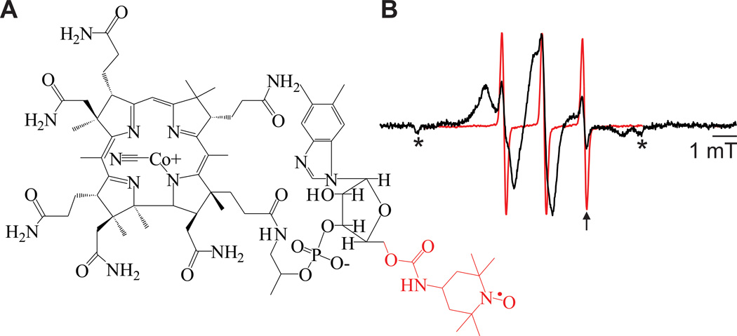
Figure 2.
TEMPO-CNCbl structure and binding. A) Structure of TEMPO-CNCbl with the TEMPO and the bonds to the 5′ carbon of ribose highlighted in red. B) RT CW EPR spectra of 10 µM TEMPO-CNCbl (red) or 10 µM TEMPO-CNCbl + 1 mM CaCl2 + BtuB reconstituted in POPC vesicles (black). The arrow indicates the characteristic resonance position for the third hyperfine line of free TEMPO-CNCbl due to its smaller correlation time and the asterisks indicate artifacts from sample tubes.

