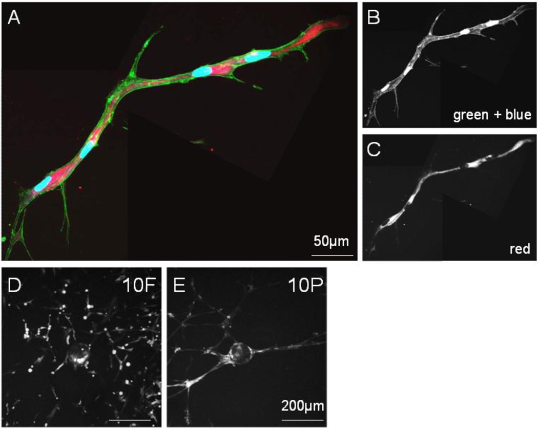
Fig. 2.
Lumen development of MSC networks in PEGylated fibrin. Two stitched frames of a single tubular structure. (A) Merged fluorescent channels from (B) and (C); (B) FITC-phalloidin labeling of f-actin filaments with DAPI staining of nuclei and (C) dextran-Texas Red localization in vesicles, scale bar = 50µm

