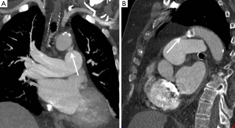
Figure 3.
Patent ductus arteriosus. (A) Coronal CT image showing a communication between the undersurface of the distal aortic arch and the left MPA branch (arrow); (B) sagittal CT image again showing the communication in the typical location for a patent ductus. CT, computed tomography; MPA, main pulmonary artery.

