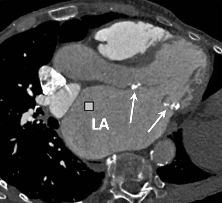
Figure 6.

Axial oblique CT image in an elderly patient with mitral stenosis secondary to rheumatic fever. The anterior and posterior mitral valve leaflets are thickened and calcified (arrows) and there is massive enlargement of the left atrium. LA, left atrium; CT, computed tomography.
