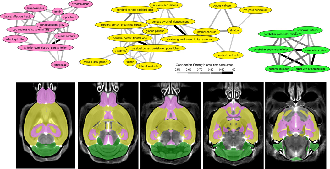
Figure 4.
Clustering of Regions - A) Bootstrapping the regions from Figure 3 revealed 3 large clusters. These clusters are connected based on the proportion of time within the same group over the 1000 bootstrapped samples. Anything above 50% was considered connected. To generalize these regions, the first (pink) cluster includes regions involved with social perception and autonomic regulation as well as some of the most sexually dimorphic regions in the brain, the second (yellow) cluster contains the majority of white matter regions, which could be representative of connectivity, and the third (green) cluster represents the cerebellar regions, which are commonly implicated in autism. B) Highlights the clusters on 5 axial slices throughout the brain and shows the interspersed nature of the pink and yellow clusters.

