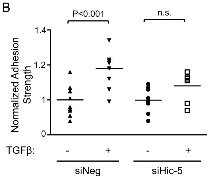
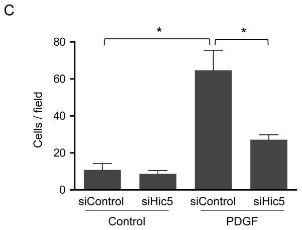
Figure 6. Hic-5 mediates TGFβ-induced cell adhesion strength and PDGF-induced migration in HASMCs.
Immediately after siRNA electroporation, HASMCs were seeded onto fibronectin-coated glass coverslips. One day post-seeding, the cells were serum-starved for 48 h, stimulated with 2 ng/mL of TGF-β for 24 h, and spun inside the spinning disk device to quantify adhesion strength, which is defined as the shear stress at which 50% of cell detachment occurs. Representative adhesion profiles for each of the each condition (A) and bar graph is the average of n=7–10 (B). Hic-5 deficient cells have impaired cells migration (C). After transfections, HASMCs were starved for 48 h, trypsinized, and reseeded in a collagen-coated Transwell insert. After stimulation for 3 h with PDGF (10 ng/mL), the cells migrating through the membrane were stained with DAPI and counted. The graph represents mean±SEM of migrated cells per field from 3 independent experiments. *P<0.01.


