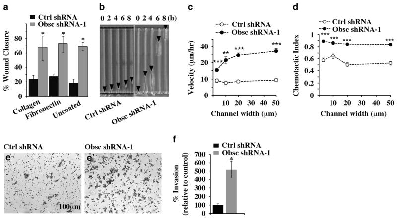
Figure 5.
Obscurin-KD MCF10A cells exhibit increased motility and invasiveness in vitro. (a) Quantification of percentage (%) of wound closure of MCF10A cells stably expressing scramble shRNA or obscurin shRNA-1 between 0 and 12 h after plating on dishes coated with collagen, fibronectin or no substrate, growing to confluency and being wounded; n =3, error bars =s.d., *P<0.03; t-test. (b) Image of control and obscurin shRNA-1-expressing cells migrating through 3-μm wide microchannels. Arrowheads point to the cell’s leading edge at the indicated time points. (c, d) Cell velocity (c) and chemotactic index (d) as a function of channel width for MCF10A cells stably expressing scramble shRNA or obscurin shRNA-1; n =3, error bars =s.e. of at least 30 cells analyzed per condition, **P<0.01 and ***P<0.001; t-test. (e, e′) Obscurin-KD and control MCF10A cells were added to a Matrigel-coated chamber and allowed to invade for 16 h. Staining of invaded cells with crystal violet was followed by quantification of the percentage of invasion of each cell population. (f) The percentage of invasion of obscurin-KD cells was compared with that of control cells, which was arbitrarily set to 100%; n =3, error bars =s.d., *P<0.03; t-test.

