. 2015 Apr 29;3(1):8–23. doi: 10.14791/btrt.2015.3.1.8

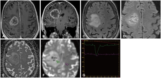
Fig. 2. 78-year-old female patient with a glioblastoma who presented with gait instability, dizziness, and dysarthria for 6-8 weeks. Preoperative MR images include axial T1 post-contrast (A), coronal T1 post-contrast (B), axial T2 FLAIR (C), axial DWI (D), axial ADC map (E), axial DSC perfusion (F), and corresponding perfusion curve (G). Post-contrast T1 images (A and B) demonstrate irregular rim enhancement and central necrosis of this tumor centered in the right frontal lobe. Surrounding the enhancing component is T2 FLAIR hyperintensity (C) which is likely infiltrative cellular edema. The lesion demonstrates restricted diffusion with low ADC values (D and E) and increased relative cerebral blood volume compared to the contralateral side (F and G) with return to baseline of the perfusion curve consistent with a primary glial tumor. ADC, apparent diffusion coefficient; DSC, dynamic susceptibility contrast-enhanced; DWI, diffusion weighted imaging; FLAIR, fluid attenuated inversion recovery; MRI, magnetic resonance image.

Fig. 2