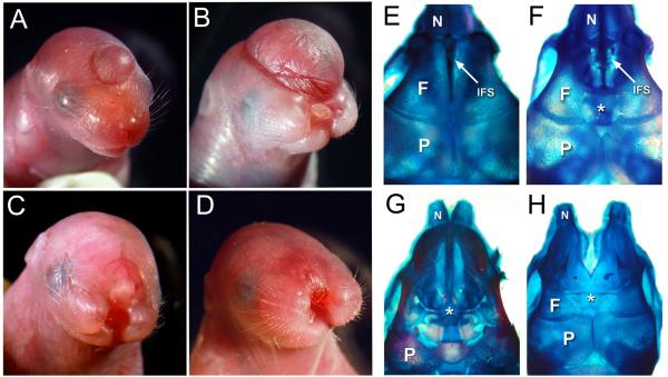
Figure 1.
Cranial traits exhibited by newborn tuft mice. (A) Anterior cephalocele. (B) Large cephalocele and midfacial cleft. (C, D) Severe midfacial cleft with an anterior cephaplocele or epidermal lesion (arrow). (E-H) Superior views of skulls indicating cranial bone (N, nasal; F, frontal; P, parietal) and suture character from a normal appearing newborn (E) compared to it’s sibling with a cyst (F) that emanated through a broadened interfrontal suture (IFS, arrow) between the frontal bones (asterisk). (G) Newborn with a bifid nose and large cephalocele similar in size to pup shown in (B), lacking frontal bones (asterisk). (H) Skull from pup with a midfacial cleft and subtle surface lesion or cyst similar to pup shown in (D), also had a broadened IFS (asterisk).

