Figure 2.
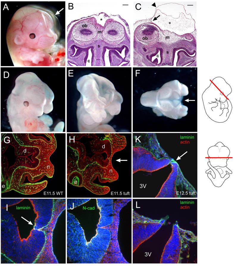
Cranial malformations in tuft embryos. (A) E14.5 embryo with a large cyst (arrow). (B) Frontal view of an unaffected E14.5 embryo and corresponding section of an (C) affected littermate with a cephalocele stained with H & E. The cyst (asterisk) extended the cranial dura meninges (arrow) and outer surface epithelia (arrowhead). Mesenchyme (m), olfactory bulbs (ob), nasal septum (s), and nasal cavity (c) are indicated. Scale bars = 200 microns. (D) E11.5 embryo with a midfacial cleft, (E) exencephaly and cleft, (F) superior view of embryo exposing anterior forebrain and midfacial cleft (arrow). (G) Corresponding horizontal sections of 3H1 wildtype E11.5 and (H) affected tuft embryos with a midfacial cleft (arrow) immunostained for laminin (green) and actin (red) at the level of the diencephalon (d), eye (e) and nasal prominences (n). Section along the embryo is shown schematically on the right. (I) Magnified view of serial sections of (H) immunostained for laminin (green), actin (red) and nuclei (blue) showing insufficient closure (arrow). (J) Serial section of (H, I) immunostained for N-cadherin (green), actin (red) and nuclei (blue). (K, L) Serial horizontal sections of corresponding levels as (H) in an E12.5 embryo with a midfacial cleft similar to (D) showing the neuroectoderm juxtaposed with the frontonasal ectoderm (arrow) via laminin (green) and exposed third ventricle (3V).

