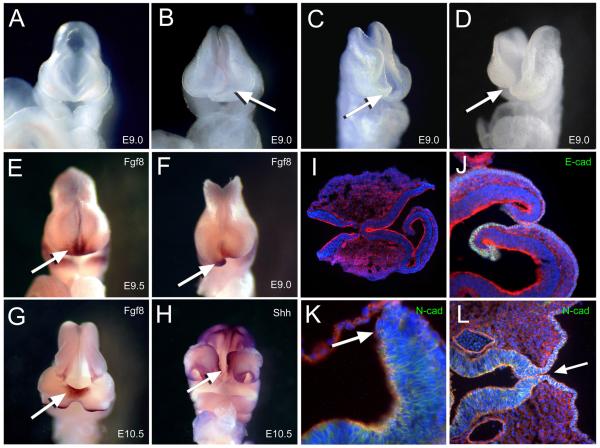
Figure 3.
Neural tube closure was affected along the anterior midline (A) Normal appearing E9.0 embryo and (B-D) spectrum of affected embryos with crooked or open neural folds along the rostral area (arrow). (E-G) Whole mount in situ hybridization of riboprobes for Fgf8 as indicated by the dense purple-brown stain marking the ANR (arrows) in (E) normal E9.0, (F) affected littermate and (G) E10.5 embryo with anterior bifidum. (H) Ventral view of embryo similar in (G) with dense purple stain marking hybridization of riboprobes for Shh along the ectoderm of the upper lip and floor plate indicating compromised rostral closure site (arrow). (I) Horizontal sections of E9 affected embryos immunostained for actin (red) and nuclei (blue) and (J) E-cadherin (green) or (K) N-cadherin (green). Arrow indicates neural tip lacking stain for N-cadherin in affected embryo. (L) Horizontal section of an E10.5 embryo similar to (H) near the neural floor plate immunostained for N-cadherin (green) indicating incomplete closure of the neural epithelium (arrow).

