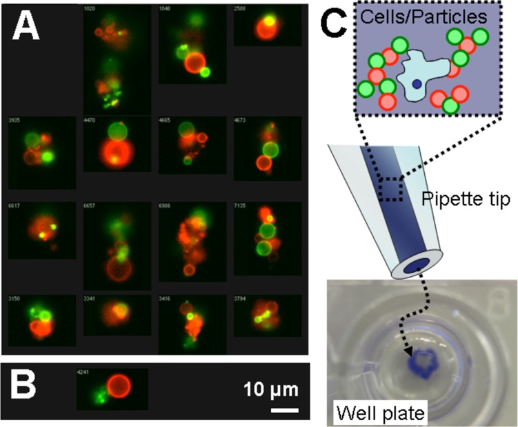
Figure 6.

Visualization of hybridized hydrogel microparticles. (A) Representative images from imaging flow cytometry show self-assembled clusters of polyacrylamide hydrogel microparticles bearing complementary DNA (green X and red X*). (B) Single micrograph of the single false-positive event from imaging flow cytometry with noncomplementary microparticles. (C) Schematic drawing shows how blue dye, cells, and hydrogel microparticles bearing complementary DNA were mixed and loaded into a pipet tip. At bottom, a digital micrograph shows the stable blue aggregate in a well.
