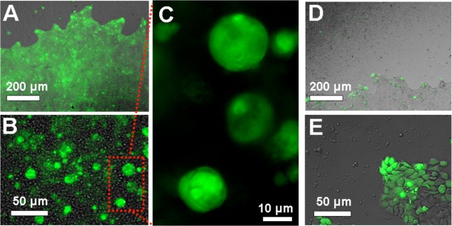
Figure 7.

Microscopy of living cells within colloidal gels composed of hydrogel particles. (A) Low-magnification brightfield/fluorescence micrograph of complementary microparticles and A431 cells. False color green overlay is fluorescence from hydrolyzed fluorescein diacetate. Scale bar is 200 μm. (B) High-magnification bright-field/fluorescence overlay image focused within the self-assembled mass. Scale bar is 50 μm. (C) Zoomed fluorescence micrograph of multicellular colonies formed within the mass. Scale bar is 10 μm. (D) Low-magnification brightfield image of cells extruded with noncomplementary microparticles. Scale bar is 200 μm. (E) High-magnification bright-field/fluorescence overlay micrograph of the edge of the confluent lawn of cells. Scale bar is 50 μm.
