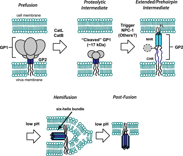
Figure 1.
Overview of GP-mediated viral membrane fusion. Upon cell attachment and uptake, the prefusion spike is first processed by CatL/CatB, leaving a 17 kDa fragment of GP1. Interaction of this remaining fragment with NPC1, and potentially other host factors, triggers the membrane fusion cascade. The GP2 fusion loop (FL) inserts into the host cell, creating an extended intermediate conformation that spans both membranes. Collapse of the N- and C-heptad repeat regions (NHR and CHR, respectively) into a six-helix bundle is promoted by low pH and facilitates progression to a hemifusion intermediate. Subsequent events lead to full fusion of both membranes. All of the steps in the fusion pathway, as well as initial cell attachment (not shown here), are susceptible to inhibition by entry inhibitors.

