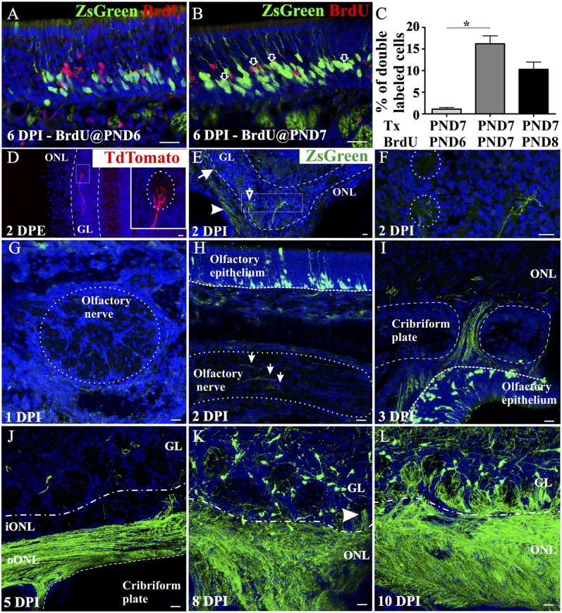
Fig. 5.
Tracking OSN axons in Ascl1CreERT2 mice. Experimental mice were injected with 4OH-Tx at PND 7 and with BrdU at PND 6 (A and C), at PND 7 (B and C), or at PND 8 (C) (n = 4 for each group). Higher levels of double-labeled cells are observed when both injections are done simultaneously (C), suggesting that BrdU and ZsGreen label the same population of OSNs within the narrow time window of basal cell division. The single asterisk indicates statistically significant (P < 0.05). Within 48 h following a postnatal electroporation of a Tdtomato-expressing plasmid, fluorescent axons can be detected branching in the glomerular layer (D). (Inset) A higher magnification of the square. Electroporation of a CreERT2 plasmid in R26RZsGreen/- mice, followed by 4OH-Tx 24 h later, shows ZsGreen expression in the outer ONL (oONL, arrowhead in E), the inner ONL (iONL, arrow in E), and the glomeruli (open arrow in E). Higher magnification of the square in E is shown in F. (G–L) ZsGreen+ axons are not observed in axon fascicles at 1 DPI (G), and only a few labeled axons are present by 2 DPI (arrows in H). At 3 DPI labeled axons cross the cribriform plate (I) and at 5 DPI (J) remain in the oONL and rarely transition into the iONL. By 8 DPI, ZsGreen+ axons enter the ventral glomerular layer (GL, arrowhead in K) and by 10 DPI innervate most ventral glomeruli (L). Draq-5 in blue. (Scale bars, 20 μm.)

