Fig. 1.
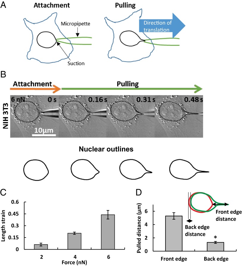
Deformation of the nucleus in a living, adherent cell. (A) Schematic of how nuclear forces were applied in a living, adherent cell. A narrow micropipette tip (∼0.5-µm diameter) was attached to the nuclear surface and forces were applied using capillary suction pressure at the contact site. The pipette was then pulled at a known rate and the nuclear response was observed. (B) DIC images show result of translation of 0.5-µm-tip micropipette sealed to the nuclear surface with a 6 nN suction force in an NIH 3T3 fibroblast cell. Translation of the micropipette pulled and deformed the nucleus as evident from the outlined shapes. (C) Nuclear deformation as quantified by length strain increased with the applied suction force (all values are different from each other at P < 0.05; n = 6 for 2 nN, n = 7 for 4 nN, n = 14 for 6 nN). (D) The deformation was much larger than the motion of the nucleus, as can be seen from the overlay of the outlines of the initial shape (red) and deformed shape (green). The extent of nuclear motion was quantified from the translation of the back edge and the extent of nuclear deformation measured as the distance moved by the front edge. The plot shows that the motion of the front edge was significantly larger than the back edge (*P < 0.05 according to Student’s t test; n = 10). Values are the mean ± SEM.

