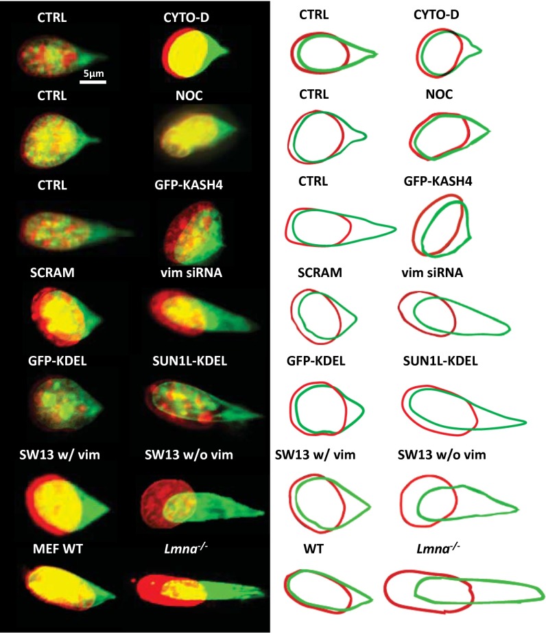
Fig. 3.
Role of the cytoskeleton in mediating nuclear homeostasis. Overlay images of a typical nucleus before (red) and after (green) forcing normal cells and in cells with perturbed cytoskeletal elements (the nucleus was stained with SYTO 59 dye; the colors are to aid visualization and correspond to the same dye). The corresponding outlines are shown on the right to aid visualization. CTRL, NIH 3T3 cells; CYTO-D, NIH 3T3 cells treated with cytochalasin-D; GFP-KASH4, NIH 3T3 cells overexpressing GFP-KASH4; GFP-KDEL, NIH 3T3 cells inducibly expressing SS-GFP-KDEL; Lmna−/−, MEFs lacking lamin A/C; NOC, NIH 3T3 cells treated with nocodazole; SCRAM, NIH 3T3 cells transfected with scrambled siRNA; SUN1L-KDEL, NIH 3T3 cells inducibly expressing SS-HA-SUN1L-KDEL; SW13 w/vim, SW13 cells containing vimentin; SW13 w/o vim, SW13 cells lacking vimentin; vim siRNA, NIH 3T3 cells transfected with vimentin siRNA; WT, wild-type MEFs.

