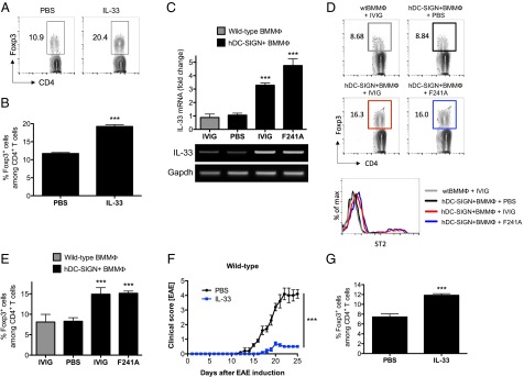
Fig. 6.

IVIG- or F241A-induced IL-33 production activates Treg cells. (A and B) C57BL/6 wild-type mice were given IL-33 (0.5 µg) i.p. on 4 consecutive days. On day 5, mice were euthanized and the percentages of splenic Treg cells were analyzed by flow cytometry. (C) Bone marrow-derived macrophages from C57BL/6 wild-type or hDC-SIGN+ mice were pulsed with PBS, IVIG, or nonsialylated F241A. Total RNA was isolated and used for quantitative real-time PCR to measure IL-33 mRNA levels. The housekeeping gene Gapdh was used for normalization. (D and E) After treatment, BMMΦs were extensively washed and adoptively transferred into C57BL/6 wild-type recipient mice. On day 5 posttransfer, mice were killed and Treg cells and ST2 expression were analyzed by flow cytometry. (F) EAE was induced in C57BL/6 wild-type mice by immunization with MOG35–55 peptide. Every 2 d postinduction, mice received IL-33 (0.5 µg i.p.). Clinical scores of disease are shown. (G) The percentages of Treg cells from draining lymph nodes of EAE mice were analyzed by flow cytometry. Means ± SEM are plotted; ***P < 0.001 determined by Tukey’s post hoc test.
