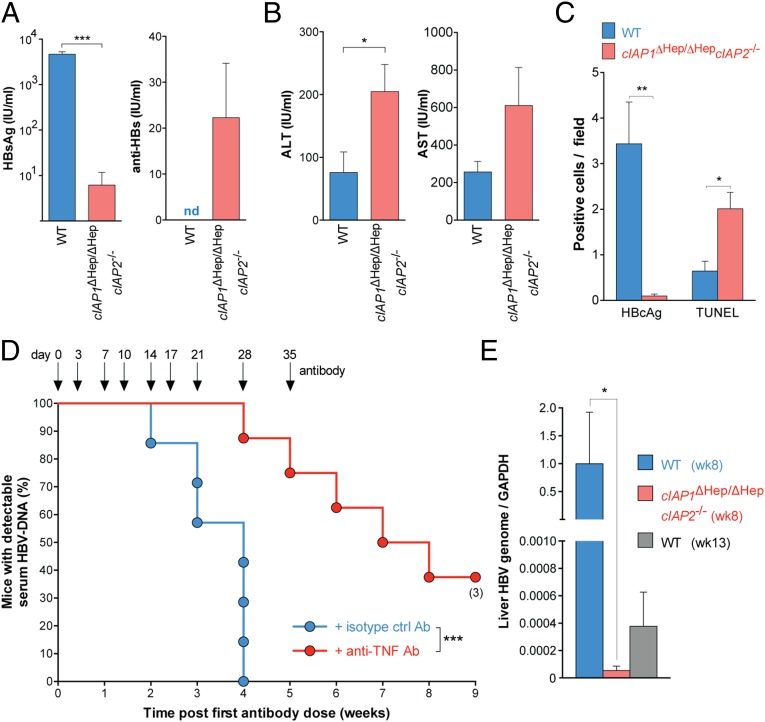
Fig. 4.
Deficiency of cIAPs promotes TNF-mediated clearance of HBV infection. (A and B) Serological HBV assays and serum transaminase levels performed 2 wk after induction of infection in mice of the indicated genotypes (n = 5 in each group). (C) Quantification of TUNEL staining and HBcAg expression in liver sections taken from HBV-infected mice 2 wk after induction of infection (n = 3–5 in each group). (D) Proportion of animals and time when cIAP1ΔHep/ΔHepcIAP2−/− mice treated with the indicated antibodies first achieved an undetectable serum HBV DNA level (n = 7–8 for each group). (E) RT-PCR of HBV DNA relative to GAPDH in the liver of infected mice with the indicated genotypes and at the indicated times (n = 4 in each group). Numbers below dots in time to event analyses represent remaining mice that have been censored. Graphs show means and SEMs, and data are representative of two independent experiments. (A, B, D, and E) Experiments were blinded. ALT, alanine aminotransferase; AST, aspartate aminotransferase; nd, not detected. *P < 0.05; **P < 0.01; ***P < 0.001 (A and B, unpaired two-tailed t test; C, unpaired two-tailed t test with Holm–Sidak correction; D, log-rank Mantel–Cox test; and E, Mann–Whitney test).

